Case Presentation: RJ is a 33-year-old mother of 2 who has been bothered by palpitations lasting 15 to 20 minutes for the last 15 years. She sometimes gets lightheaded and nearly passed out in a grocery store while holding her infant son. She was diagnosed with SVT (supraventricular tachycardia) and was given different medications that either produced severe fatigue or did not prevent episodes. She was told there were other treatment options but her doctor wasn’t sure which was best for her. He referred her to an electrophysiologist (heart rhythm specialist).
In the last 15 years, the treatment of individuals who suffer from episodes of abnormally rapid heart rates (tachycardias) has undergone tremendous changes. New drugs and implantable devices (pacemakers and defibrillators) have been part of this process, but among the more dramatic developments has been the use of a heart catheterization technique called catheter ablation (CA). The heartbeat is the result of heart muscle cells transmitting an electrical signal from one cell to another. When this transmission becomes abnormal, the heartbeat can race inappropriately. Using the CA technique, the electrophysiologist can determine the exact location of the abnormal heart muscle and eliminate (ablate) it. The procedure is applicable to a wide variety of tachycardias, has an extremely high success rate for many of these, and has a low complication and rate of tachycardia recurrence. CA works by delivering energy through an electrode at the end of a catheter that has been threaded through a vein or artery to the heart. The most common type of energy used is radiofrequency (RF), which coagulates the abnormal tissue that is in immediate contact with the electrode. A small scar develops at this site, and the effect is usually permanent. Other types of energy, such as microwave, sonar, or cryo (freezing) can be used, but the most common application is with RF.
What Types of Problems Are Treated by Catheter Ablation?
CA can be used to treat a wide variety of tachycardias, which can involve heart tissues in the upper chambers (atria), also called supraventricular, or the lower chambers (ventricles), called ventricular tachycardias (VT). In many cases, a small number of cells (a "focus") start firing rapidly and CA can eliminate the focus (Figure 1). In other types of tachycardia, an electrical circuit exists within which the electrical signal travels more or less in a circle ("reentry"). Most cases of SVT and VT are due to reentry. A special type of SVT, called atrial fibrillation, is characterized by extremely fast impulses in the atrium (up to 600/min), for which CA can be used either to decrease the number of impulses getting to the ventricles or in some cases to locate and ablate the area(s) from which the fibrillation starts.
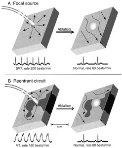 Figure 1. How catheter ablation works in different types of tachycardias. Sections of heart muscle where tachycardia originates are depicted. A, A focus (*) is repeatedly firing to cause tachycardia by spreading outward to other cells; the ECG below shows SVT at 200 beats/min. A catheter with a large electrode at its tip is in contact with the focus; after ablation (right), the area of the focus has been damaged and is no longer firing. Arrows show normal electrical propagation and the ECG below is normal. B, A reentrant circuit is shown with electrical propagation around a nonconducting barrier to produce VT at a rate of 180 beats/min, as shown in the ECG below. The impulse must pass through a narrow "bottleneck" to continue. This is an area (where catheter tip is pointing) at which a small amount of damage can eliminate reentry; after ablation, the bottleneck is sealed off, preventing reentry. The ECG rhythm below is normal.
If I Have Tachycardia, Does That Mean I Have "Heart Disease"?
Not necessarily; in fact, in the majority of individuals under 45 years old who have tachycardia, the disorder has nothing to do with structural heart disease such as valve or coronary artery problems. Almost all SVTs and a few VTs are of this relatively benign type and rarely cause death or other serious consequences. However, some tachycardias, such as most types of VT, do occur in people whose hearts are damaged (heart attack or cardiomyopathy [weak heart muscle]). In these cases, the rhythm disturbance is much more dangerous and can cause sudden death.
What Goes on Before and During a Catheter Ablation Procedure?
Once a CA is scheduled, patients usually stop taking medications previously used to try to control the tachycardia because on the day of the procedure, the electrophysiology (EP) doctor often needs to start an episode of tachycardia to determine what type it is and where it is coming from. After discussing risks and benefits of the procedure and giving informed consent, the patient is brought to the EP laboratory. Local anesthetic is used before catheter insertion in the groin and perhaps in one side of the neck, the elbow, or the area under the collarbone; 3 to 5 catheters are often used simultaneously. Once catheters are in the veins or artery, fluoroscopy (x-ray) is used as they are moved toward the heart and positioned in strategic locations. Recordings of the heart’s electrical activity are made from inside the heart. After tachycardia induction, a special ablation catheter is then maneuvered so its tip electrode is in contact with the abnormal tissue. The location of this ablation target is determined by a process called mapping, in which the ablation catheter is moved from spot to spot until the appropriate area is found. At this point, RF energy is turned on and, if the catheter location is correct, the tachycardia is eliminated (Figure 2). Testing is performed to see if tachycardia can be initiated again and the procedure is repeated if necessary. If tachycardia cannot be reinitiated, catheters are withdrawn from the body. The procedure usually takes 2 to 4 hours. In experienced laboratories, most types of SVT can be cured in over 95% of patients. VT in patients without structural heart disease is curable more than 90% of the time, whereas in patients with structural heart disease (for example, prior heart attack), the complete cure rate is lower, only 40% to 50%. In these patients, almost all of whom also receive an implantable defibrillator, CA is used to decrease the number of times the defibrillator is used, rather than as a cure from all VT episodes. For more information on cardioverter-defibrillators, see the Circulation Cardiology Patient Page by Reiffel and Dixon (Reiffel JA, Dixon J. The implantable cardioverter-defibrillator: patient perspective. Circulation. 2002;105:1022–1024).
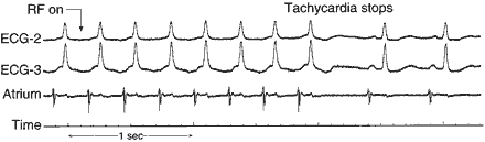 Figure 2. Ablation of an SVT. Two electrocardiographic recordings (ECG) and a recording from a catheter in the atrium are shown during an episode of SVT at 230 beats/min. Radiofrequency (RF) energy is delivered at a spot chosen by mapping. Tachycardia stops suddenly and normal rhythm resumes after 2 seconds of ablation energy.
"Damaging Tissue": Isn’t That Dangerous?
The amount of heart tissue that is damaged during a typical CA procedure (3x5 mm) is insignificant for overall heart pumping function. The scar tissue formed appears to remain stable over time and to not cause problems even years later. Nonetheless, things can go awry during the procedure. Possible complications vary with the specific tachycardia diagnosis, but among these are perforation of the heart with leakage of blood into the sac surrounding the heart, perforation of a blood vessel with leakage outside it, inadvertent interruption of normal conduction (which requires a pacemaker), stroke, heart attack, and even death. These are all very rare. A pacemaker is needed in less than 1 in 200 cases, and other, more serious complications occur in less than 1 in 500.
What Happens After the Procedure?
Patients are observed for a few hours for symptoms, rhythm problems, and bleeding from the catheterization sites. In many cases, at the end of this observation period, they may be discharged. Others stay overnight in the hospital. Aspirin is often prescribed for 2 to 4 weeks to minimize risk of clot formation at ablation sites. Patients can perform light activity (walking, stairs) almost immediately in most cases, with resumption of full work or school schedules within a few days. A follow-up visit to the EP doctor is often useful to check the catheterization sites and review the procedure.
Are There Alternatives to Catheter Ablation?
Medications can be very effective in some, but for many patients, the cost, inconvenience, side effects, and often-poor efficacy make them less attractive. For some patients, implantable pacemakers and defibrillators are good or even preferred options.
Who Are Candidates for Catheter Ablation?
Because of the very high success rates and low recurrence and complication rates for many types of tachycardia, CA is very reasonable therapy for most individuals who have enough problems with tachycardia that they require some form of treatment. It is particularly worth considering when medications have failed to control episodes. Our patient at the beginning of this article would thus be an excellent candidate. For those individuals with very rare episodes that cause minimal symptoms, CA may have little to offer; however, these comprise a minority who present to an electrophysiologist.
In summary, CA has become a major tool in treatment of tachycardias, with a wide applicability to a variety of tachycardias, a very high success rate, and a very low incidence of complications. For individuals who are bothered enough by episodes of abnormally racing heart that they need some type of treatment, CA is an excellent option.
Footnotes
Drs Miller and Zipes consult with Medtronic, Inc, regarding ablation technologies.